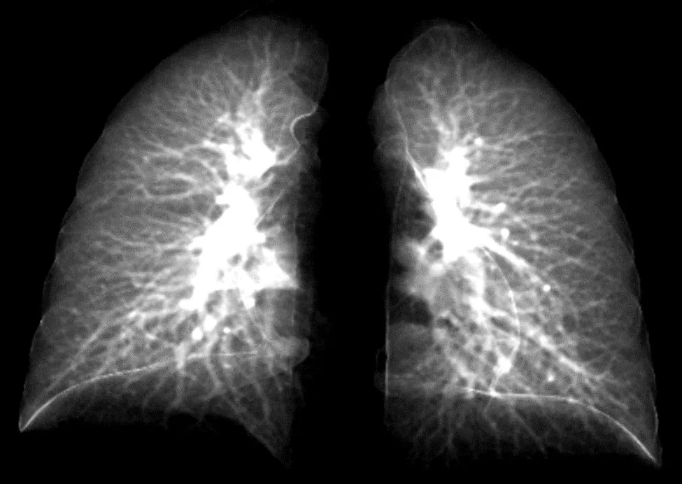
College of Iowa researchers have created a sophisticated mannequin that may detect lung harm in long-COVID sufferers utilizing a easy chest X-ray. The mannequin takes information factors from 2D lung pictures constructed from 3D CT lung scans. This picture reveals particulars of the lung in a 2D picture. The mannequin is engaging, as a result of 2D chest X-ray gear is extra generally obtainable and less expensive. Credit score: Ching-Lengthy Lin lab, College of Iowa
Scientists create a brand new mannequin to detect COVID-19’s effects using 2D chest X-rays.
For patients dealing with lingering respiratory symptoms from COVID-19, a traditional chest X-ray can reveal only so much. The two-dimensional (2D) scans simply can’t distinguish compromised lung function resulting from the novel coronavirus. For that diagnosis, a more expensive, three-dimensional (3D) technique called a computerized tomography (CT) scan is necessary.
However, many medical clinics in the United States don’t have CT scanning equipment, leaving so-called long-COVID patients with little information about their lung function.
That may soon change. In a new study, something called a contrastive learning model has been developed by scientists at the University of Iowa. This model “learns” how to detect compromised lung function in long-COVID patients using composite 2D images constructed from 3D CT images. Another technique, called transfer learning, then conveys lung diagnostic information from a CT scan to a chest X-ray, thus allowing chest X-ray equipment to detect abnormalities the same as if those patients had used a CT scan.
In the study, which was published recently in the journal Frontiers in Physiology, the researchers showed how their contrastive learning model could be applied to detect small airways disease, which is an early stage of compromised lung function in long-COVID patients. Of the long-COVID patients, the models were advanced enough to distinguish the severity of the compromised lung function, separating those with small airways disease from those with more advanced respiratory issues.
“The new element to the model is taking information from 3D CT scans showing lung volume and transferring that information to a model that will show these same characteristics in 2D images,” says Ching-Long Lin, Edward M. Mielnik and Samuel R. Harding professor and chair of the Department of Mechanical Engineering in the College of Engineering at Iowa. “Clinicians would be able to use chest X-rays to detect these outcomes. That’s the bigger perspective.”
The researchers based their modeling on CT scans of 100 people who were infected with the original COVID strain and went to UI Hospitals & Clinics for diagnosis for breathing problems between June and December 2020. Many of these long-COVID patients had small airways disease, a diagnosis reported by Alejandro Comellas, clinical professor of internal medicine–pulmonary, critical care, and occupational medicine, in a paper published last March in the journal Radiology.
Small airways disease affects a network of more than 10,000 tubes at the nexus in the lung where oxygenated air mixes with blood to be carried throughout the body. People with small airways disease have many of these vessels constricted, thus limiting the oxygen-blood exchange in the lungs, and impeding breathing overall.
Lin and his team collected data points at two intervals in the CT lung scans—when the patient inhaled and when the patient exhaled. The researchers compared their results with a control group that had not contracted the virus as they created the contrastive learning model.
“Our models successfully identified decreased lung function from long-COVID patients compared to those who had not gotten the virus,” says Lin, whose expertise is in machine learning and computational fluid and particle dynamic simulation.
Lin’s team advanced the model so it could separate patients with small airways disease from those with more advanced complications, such as emphysema.
“The study demonstrated in an independent way that patients with post-COVID have two types of lung injuries (small airway disease and lung parenchyma fibrosis/inflammation) that are persistent after having recovered from their initial SARS CoV-2 infection,” says Comellas, a co-author on this study.
“Chest X-rays are accessible, while CT scans are more expensive and not as accessible,” Lin adds. “Our model can be further improved, and I believe there is potential for it to be used at all clinics without having to buy expensive imaging equipment, such as CT scanners.”
The authors note the study is limited, in part because the sample size is small, and the patients are from a single medical facility. A larger sample size, they write, may uncover more variations in lung function stemming from long COVID.
Reference: “Contrastive learning and subtyping of post-COVID-19 lung computed tomography images” by Frank Li, Xuan Zhang, Alejandro P. Comellas, Eric A. Hoffman, Tianbao Yang and Ching-Long Lin, 11 October 2022, Frontiers in Physiology.
DOI: 10.3389/fphys.2022.999263
Co-authors, all from Iowa, include Frank Li, Xuan Zhang, Eric Hoffman, and Tianbao Yang.
The National Heart, Lung, and Blood Institute, a branch of the U.S. National Institutes of Health; and the U.S. Department of Education funded the research.
#LongCOVIDs #Results #Detected #Easy #Chest #XRays
Source